


The forearm is the site of the upper limb,It consists of two bones and is bounded by two joints. Often students confuse the location of the shoulder and forearm in the anatomy of a person. In order to understand, consider the structure and functions of the portion of the upper limb on the forearm.
On average, the schools begin to study the coursehuman anatomy. This is a very interesting and at the same time an extensive section of biology, which requires a qualitative assimilation of knowledge. Human anatomy is a science that considers the structure of an organism, its functions and vital activity in general.
About where the forearm of a person is,tell a skeleton model or anatomy atlas. Having carefully studied the name, location, types and purposes of bones, it is easy to understand the structure of the upper limb of a person - the hand.
Mobility of the skeleton is provided by the muscles thatattached to the bones. Considering the forearm, where there are many different types of muscle tissue, you can understand how the hand comes into motion. The contraction of muscles is facilitated by nerve impulses, which are taken by the nerves located in the forearm.
So, a bit of theory. On the question of where the forearm is located in person, anatomical atlas or skeleton mock-up will help. The forearm bones are one of the components of the upper limb of a person, playing an important role in the mobility of the hands. Having carefully studied the image of the skeleton, it is easy to see where the shoulder and forearm are.

From the name you can guess that the forearmis located below the bones of the shoulder, it precedes it and together forms the basis of the upper limb of man. In order to understand the anatomy of the upper limb, it is necessary to study the bones that make up it.
Part of the upper limb is the forearm, wherethere are two bones, has a fairly simple structure. His tubular long bones are made up: the elbow and the ray, each of which has its own structural features.
What you need to know about the structure of tubular long bones:

The body of the ulna and radius has a triangularform, which means the presence of three surfaces. The front part of the bone is facing forward, the back is facing back. The location of the third side of the radial and ulnar bone varies.
We have already found out that the forearm is wherethere are two long tubular bones connected on both sides with joints. The third side of the ulna is called the medial and is turned inward, as are its edges.
The ulna is located in the outer part of the arm,that is called the medial position. For example, if we consider the limb from the left and right sides, then the left forearm contains the ulna bone on the left, and the right forearm on the right. In other words, the ulna is in the forearm from the little finger.

Upper epiphysis of the ulna thickerproximal epiphysis of the radius and articulated with it using a block-shaped notch, which is restricted by two processes: coronary and ulnar. From the inside of the coronoid process there is a radial notch, intended for the head of the radius. The joint in conjunction with the articular surface and cartilage forms an elbow joint, ensuring flexion and extension of the forearm.
The lower epiphysis, on the contrary, is thinner than the distal epiphysis of the radial bone and connects to it with the help of an articular circle, and then passes into the wrist joint.
If the ulna is located medially, thenRadial third is not given: it is located on the inner side of the hand, ie, it is located distally. For example, the left forearm (where the ulnar bone is located on the left) contains the radial bone on the right. In other words, the radius is located on the side of the thumb.
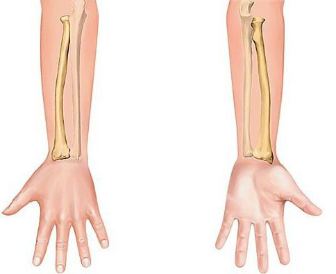
The third side of the radius is calledlateral, it is turned outward. The upper epiphysis consists of a head with a small indentation in the center, intended for connection with the condyle of the humerus. The distal epiphysis contains the ulnar incision from the outside for connection to the ulna head.
Important role in the anatomy of tubular bonesThe forearm plays a way of connecting them together. The joints provide movement of the radius around the ulna. The bone can move toward the inner or outer side, while the wrist and elbow joint always act together.

When making a motion, the ray describesan arc of 140 degrees of the ulna. At the same time, the brush and shoulder are shown, albeit in slight motion, that in the total amount from 220 to 360 degrees of the volume of movements. It is the possibility of such rotation that allows a person to make various movements of the upper limbs.
Important place in joining the bones of the forearmoccupies the interosseous membrane, consisting of collagen fibers. It is located between the ridges of the radial and ulnar bone and keeps them in such a way that it does not constrain movements.
The people's understanding of the locationshoulder introduces confusion at the beginning of the study of human anatomy. The fact that everyone is accustomed to be considered a shoulder, in medicine is called a shoulder girdle, or a shoulder-strap. Then where is the shoulder, and where is the forearm? The shoulder is the portion of the upper limb from the shoulder to the elbow joint, through which the humerus is connected to the bones of the forearm.

The shoulder is connected to the lateral radiuspart of the articular surface, which has the form of a ball. This is the head of the humerus of the humerus. With the ulna bone, the shoulder is joined by the medial part, forming a block of humerus. The venous and ulnar processes enter the block in front and behind, respectively. Above the block there are pits, in which, when bending or unbending the elbow, there are appendages.
Where a person has a forearm, we have already figured out. Let us consider in more detail which muscles are involved in this segment of the upper limb. Depending on the movements, the muscles of the forearm can be divided into:

The named groups of muscles unite in two basic categories depending on their position: front and back.
The front muscles, which move the forearm,- this is where the radial bone turns inward. The group includes muscle flexors. The superficial layer of the muscle tissue of the anterior muscles begins with the medial part of the epicondyle of the shoulder. Deep muscle layers begin on the bones of the forearm and the membrane between them. The perforators are attached to the radius.
Muscle tissue is closer to the humerus, more pronounced, and near the wrist band is mainly represented by tendons.
The superficial layer of the anterior muscles is:
The deep layer of anterior muscles is represented by a longflexor of the thumb, deep flexor of the fingers and a square pronator. The forearm, where there are many muscles involved in the movement of the upper limb, determines the dexterity and variety of actions.
Back muscles of the forearm are the arch supports and extensors. The superficial layer of muscle tissue consists of muscles:
The deep layer of the posterior muscles of the forearm is represented by the arch support, the long muscle (participates in the lead of the thumb), the short and long extensor of the thumb and the extensor of the forefinger.
The forearm, where there are many muscles, is also the place of passage of nerves. Let's consider the basic nerves located in this area, and their functions:
The bones of the forearm are thin enough, so that they easily break down even with small injuries.

Fractures of the forearm can be divided into several types:
In case of fractures, it is extremely important to provide first aidthe victim. If the fracture is open, do not attempt to fix it yourself. It is necessary to remember that two joints are necessarily immobilized: wrist and elbow. To do this, you can impose a tire, without touching the side of the hand from which the bone protrudes.
The forearm is an important segment of the upper limb, providing high mobility of the hand and a variety of performed actions.
</ p>
